- 01 Overview
- 02 CMMS Research Track Record
- 03 How it works: CMMS and muSR
- 04 How it Works: CMMS and bNMR
01 Overview
TRIUMF’s Centre for Molecular and Materials Science (CMMS) equips Canadian and international researchers with unique particle beam tools to explore materials’ atomic-level details and guide the way to next-generation technologies and medicines.
The CMMS provides two powerful research tools: Beta-detected Nuclear Magnetic Resonance (bNMR, the Greek-letter b is pronounced ‘beta’); and Muon Spin Resonance (µSR, pronounced “mew-ess-are”). CMMS is North America’s only µSR facility and one of four worldwide.
As researchers develop new materials and new applications for existing materials, it’s necessary to understand and characterize materials’ atomic-level characteristics. While other techniques can reveal atomic-level structure, µSR and βNMR provide information on otherwise invisible atomic-level qualities: magnetic and electric fields; and electron behaviour. It’s these qualities that often determine whether a material will make a breakthrough battery, a new superconductor or cutting-edge computer chip.
Both bNMR and µSR involve implanting a radioactive probe, whether muons, or in bNMR the rare isotope lithium-8 (8Li), into a gas, liquid or solid. The muons and 8Li are microscopic spies, picking-up information about a material’s atomic-level properties and rapidly communicating this out to scientists via their decay products.
The techniques provide both the highest sensitivity to atomic-level magnetic and electrical details and numerous other unique advantages. µSR can be performed with tiny samples, less than the size of a dime, enabling researchers to test new materials in very early R&D stages when only small quantities can be produced, and thus it’s often one of the first tools used in analysis. In bNMR, 8Li can be precisely implanted with nanometer accuracy making bNMR exceptional at the study of the localized magnetic and electronic properties of ultra-thin films, nanostructures and interfaces. 8Li has a half-life of 1.2 seconds, 500,000 times longer than that of a muon, so bNMR and μSR can be used to study phenomena over a wide range of time scales.
Each year about 100 Canadian and international scientists bring their material samples to CMMS for testing, notably in magnet and superconductor research. Commercial and academic users collaborate with CMMS’ ten scientific and technical staff who provide expert guidance and input on experimental design to maximize CMMS resources. Similarly, CMMS in-house research group has a scientific program that both attracts new users through demonstrating the power of bNMR and µSR as research tools and providing opportunities for collaboration.
02 CMMS Research Track Record
03 How it works: CMMS and muSR
Located in the Meson Hall, the μSR facility allows users bring their own research samples to CMMS; usually new or rare materials, for example nanotextured metal alloys for magnets or superconductors. µSR academic users apply through a competitive process overseen by an eight-person international expert scientific committee that assesses the feasibility and scientific interest of the proposed experiment. Academic users have free access to CMMS facilities; commercial users, those whose research results are not published in the open literature, pay $15,000/per day for use of the CMMS facility.
The physics
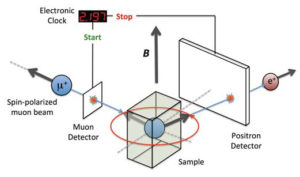
µSR is a much more sensitive variant of the better known nuclear magnetic resonance (NMR), one of the most important tools in science for characterizing substances. µSR uses beams of polarized, positive muons (μ is the Greek letter “mu”) that are implanted into a material coming to rest in the space between atoms. There, a muon’s spin is changed by the local magnetic environment. Each muon’s spin precesses, or wobbles like a top, around the local magnetic field in the material.
The positive muons have a half-life of just two millionths of a second and decay into positrons (anti-electrons) which are emitted in the direction of the muons’ spin. Since the implanted muons had a known polarization, by observing the directions in which the positrons are emitted scientists can determine how the internal magnetic fields have affected the muons’ spins and reconstruct these magnetic fields.
Muons can also be used to study chemical processes. A muon’s mass is about a one-ninth that of a proton and thus a positive muon behaves as a tiny version of a proton. When positive muons are implanted into a material they pick up an electron and form muonium (Mu), a variant of hydrogen. Chemists study the reactions of Mu with different chemicals and in different environments and also use Mu to create free radicals and study the structure, motion and reactivity of these short-lived and highly reactive compounds. Using Mu, chemists are looking at a range of questions from fundamental ones about how mass affects reactions rates to more applied ones such as how antioxidants scavenge the free radicals that are responsible for diseases and aging.
The μSR facility consists of two main components: the muon beamlines; and the muon spectrometers and associated cryogenic and heating units.
Making muons and muon beamlines
In the Earth’s upper atmosphere muons are naturally created by the interaction of cosmic rays with gas molecules. At TRIUMF, a muon beam is created by firing 500 MeV protons into a graphite target to produce positive and negative pions. The pions decay in about 26 billionths of a second into a muon and a muon neutrino. The positive pions all decay into positive muons that are spin-polarized, all muon spins are pointing in the same direction. The level of spin polarization is much higher than conventional magnetic resonance techniques and is a main reason that μSR is more than a billion times more sensitive than NMR.

Presently, muons are supplied to experiments via two muon beamlines, M15 and M20. In 2019 two additional beamlines will be added: one supplying higher energy muons for experiments at high pressures, low temperatures and in strong magnetic fields; and the other specialized for full-support, fast-turn-around experiments for users new to μSR in which CMMS staff will provide full support from experimental design, running the experiment, analysis of the data and writing up the results.
Muon spectrometers
The test sample is placed in the centre of a muon spectrometer surrounded by a magnet. An incoming muon passes through a muon counter, which starts a fast-electronic clock that is stopped by the detection of the corresponding decay positron. The muons are implanted about one millimetre into a sample with their spin either parallel or perpendicular to an external magnetic field created by a surrounding magnet.
μSR includes a suite of eight different spectrometers and related heating and cooling equipment that enables experiments to be conducted over an extremely wide range of temperatures, from just 0.015 oC above absolute zero (-273 oC) to more than 900 °C and over a wide range of magnetic strengths.
For example, the Helios spectrometer has a powerful superconducting magnet that creates a magnetic field up to six Tesla providing the ability to scan, or make step-wise, measurements of material properties over a wide range of magnetic field strengths. Alternately, the LAMPF spectrometer can operate with zero magnetic field, making it ideal for studying magnetic materials. The Dilution Refrigerator spectrometer operates at the very low temperatures required for the study of superconducting materials such as niobium. Each spectrometer has a distinct physical design and is supplied by one or more different cryostats or ovens for cooling or heating samples.
04 How it Works: CMMS and bNMR
bNMR is a next-generation form of NMR– a billion times more sensitive than NMR – used to probe atomic-level magnetic fields and the way that these fields change.
The higher-resolution view bNMR provides is critical in the current search for superconductors and nanomaterials and to better understand complex, biologically important molecules. For example, scientists need to characterize the special electrical or magnetic properties at material surfaces, or at the metal ion binding sites around which biomolecules fold.
bNMR’s high level of sensitivity comes from its combination of a high percentage of both isotope polarization and beta particle detection.
Beta decay (the beta in bNMR) is a form of radioactive decay in which an unstable nucleus becomes more stable by emitting a fast, high-energy electron: a beta particle.
An intense, pure beam of 8Li or 31Mg is produced in the ISAC target facility and sent via beamline to the Laser Spectroscopy facility where the rare isotopes, in flight, are highly polarized, their nuclei made to all spin in the same direction. The nuclei continue via beamline to the bNMR facility where the polarized beam is fired into the test material at just the right energy to come to rest where they can provide information that’s of interest to researchers. TRIUMF researchers have demonstrated the ability to control the implantation depth in solids to just 2.5 nanometers or about 25-times the size of an atom.
Since the 8Li nuclei are polarized the researchers know how they are oriented when implanted into the sample. Once at rest, each embedded isotope is affected by the magnetic characteristics of its local atomic environment, which in turn affects the spin and direction of emission of the beta particle. The beta particle therefore carries a detailed characterization of the local magnetic fields within the substance, which is recorded by the bNMR’s sensitive beta particle detectors.
TRIUMF scientists have demonstrated that the bNMR facility can effectively measure novel detail about nano-level electromagnetic characteristics at surfaces and interfaces. This includes structure, phase transitions and barrier dynamics. This makes bNMR a powerful new tool for optimizing functionality in novel nanostructures and identifying materials with localized novel magnetic or superconducting characteristics. This ability is particularly important with the further miniaturization of electronic components, such as transistors, where electronic and magnetic phenomena at surfaces and interfaces play a larger role in overall functionality.
bNMR and TRIUMF Life Sciences
TRIUMF’s Life Sciences researchers are pioneering the use of bNMR in characterizing the role of metal ions in biologically important biomolecules. Many biomolecules, including chlorophyll, RNA, insulin and beta-amyloid have structures that fold around key metal ions, including magnesium, copper and zinc.
In bNMR, a radioactive metallic isotope is incorporated into a biomolecule’s structure to act as an incredibly sensitive built-in probe transmitting otherwise unattainable information. This is done through the beam implantation of radioactive metallic isotopes, including 31Mg, into solutions of target molecules.
TRIUMF’s bNMR offers the first opportunity to study localized structure and dynamics around metal ions in biomolecules. This major advance is facilitated by:
- TRIUMF’s bNMR researchers have provided the proof-of-principle that beam embedded 31Mg isotopes become functionally bound in biological samples. This bNMR achievement opens a new scientific frontier in the detailed characterization of metal-ion related protein structure and dynamics.
- The invention of a new, patented liquid-phase bNMR spectrometer optimized for the study of biological samples. The new bNMR spectrometer will provide an unprecedented perspective on liquid samples at biologically-relevant temperatures. In conjunction with the existing bNMR spectrometers, the new spectrometer will provide for triple data-taking within one experiment.